
The elderly Eskimo woman moved aside the notched stones which held down the skin cover on the sod roof and climbed down into her semi-subterranean house. Although it was a cool day, the heating-cooking fire quickly warmed the little room. She was able to take off her clothes, soaked from wading in the tidal pool where she gathered seaweed and shellfish. This ability to obtain food from the sea made life relatively easy here, even for a widow whose children had long since grown up and now lived with their own families. Although fuel was something of a problem for everyone on the small island, in what was later to be called the Bering Sea, she was able to gather enough driftwood to keep her fire going. The men were generous with the walrus blubber that made up the dietary staple. As the permanently frozen ground made it impossible to grow anything, the sea was the source of life for the Eskimos.
The naked old woman sat by her fire, stirring her bubbling soup and coughing slightly. The smoke from the fire seemed to bother her more than it used to, but she was happy not to feel the pain in her chest that frightened her so. Except for these intimations of mortality, it was a snug little world. Within seconds that little world ended. Without warning the earth trembled and cracked. The air filled with moss as the roof of her house disintegrated. Her scream was stifled by the moss that filled her lungs, and consciousness was lost when one of the stones from the roof hurtled down and struck her forehead. She fell into one of the cracks in the floor, where she was quickly covered over by the debris of the house and suffocated.
By the time the surviving villagers got to her house, there was nothing they could do. Rather than find and clothe her now frozen body for proper burial, they attempted to reconstruct their own lives, shattered by an earthquake which had lasted only a few seconds.
Sixteen hundred years later, in September of 1973, a group of scientists from the Universities of Pennsylvania and Alaska, the National Park Service, and the Arctic Health Research Center of Fairbanks, Alaska, stood around the still-frozen body of this woman, at the Research Center. Beach erosion on Cape Kielegak, St. Lawrence Island, had allowed her long-buried frozen body to tumble from a cliff to the underlying beach, where she had been found by passing Eskimos. They reburied her body in the permafrost and notified the National Park Service. Zorro A. Bradley of the Park Service arranged for the body to be shipped by air to Fairbanks, where it fell under the jurisdiction of the coroner, Mr. Lincoln E. Ost. He obtained a court order for an autopsy. As it did appear likely that this was an ancient body, the Anthropology Department of the University of Alaska was also notified. They contacted Dr. J. Lawrence Angel, Curator of Physical Anthropology at the Smithsonian Institution, and Dr. Angel, knowing of my interest in mummies, referred them to me. Thus I was able to perform this unique autopsy, the results of which, combined with archaeological data, led to the above reconstruction.
The findings of the autopsy were as follows. The body, radiocarbon dated at A.D. 400, was almost perfectly preserved. There were no clothes, which one would have expected to be equally intact. Eskimos are unclothed only in their houses, customarily being clothed for deliberate burial. The air passages in her lungs were packed with moss, and microscopic examination revealed hemorrhage, which is associated with suffocation. Her lungs also showed anthracosis (an accumulation of carbon pigment), and emphysema, accounting for her chronic cough. This disease in modern patients is associated with cigarette smoking or air pollution, but when seen in ancient bodies is due to lifelong exposure to the smoke of open cooking and heating fires in poorly ventilated houses. Her chest pain can be attributed to a moderate degree of coronary arteriosclerosis, although there was no evidence that she had suffered a myocardial infarction (“heart attack”).
One of the aims of paleopathological investigations is to provide such reconstructions of the life of individuals in the past. Disease and death are integral parts of the history not only of individuals but of whole populations. The evolution of diseases and their role in human history are the subjects of paleopathology, the study of the diseases of ancient peoples.
Information on diseases in the past can be acquired through several sources. At present, our concept of disease acting on the population level is primarily based on historical records, such as the Egyptian medical papyri, or records of the great plagues of medieval Europe. Another example is the role of epidemic diseases, such as measles, smallpox and yellow fever, in the decimation of the Indian population of the New World after European colonization. Contemporary writings and census figures are useful in these studies, although somewhat limited by the difficulty of applying modern diagnostic terms to descriptions several hundred years old.
Works of art often give information on diseases; these include such diverse sources as paintings, pottery effigies, and figures and faces on coins. However, the most reliable information comes from examination of actual pathological specimens, including both skeletal material and mummies.
Most disease processes leave little or no mark on the bones. Mummies, which are defined as bodies preserved either naturally, by freezing or drying, or artificially, hold a much greater potential for paleopathological examination. Artificial mummies are found not only in Egypt but also in Peru, the Aleutian and Canary Islands, and the islands of the Torres Strait, off Australia, Frozen bodies have been found in the mountains of Peru as well as in Alaska, while the southwestern United States is a rich source of desiccated mummies. Sufficient numbers of mummies exist that such studies can be expanded to the population level. My studies of these various types of mummies have led to field work in such diverse areas as Alaska and Egypt, and will eventually constitute my doctoral thesis in anthropology.
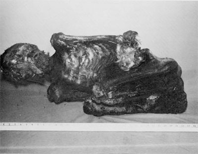
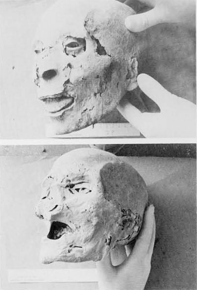
The first mummy I studied represented an interesting combination of natural and deliberate mummification. The individual was an Aleut, a people inhabiting the Aleutian Islands and only distantly related to the Eskimos. He had died in the early 18th century A.D.; the Aleuts had been mummifying their dead for only a few decades before the Russians’ arrival in 1740 completely disrupted the native culture.
It is felt that the mummification techniques used by the Aleuts were based on the anatomical knowledge acquired from butchering sea mammals, a knowledge attested to by an extensive anatomical vocabulary. The procedure used varied with the social status of the deceased. Tribal leaders were eviscerated through an abdominal incision, the body cavities stuffed with grass, and the bodies dried for several weeks before being wrapped in several layers of animal skins. Those not accorded this special treatment were simply wrapped up, and all of the bodies were placed in a burial cave on Kagamil Island, in the mid-Aleutian chain. A volcanic vent in this cave dried out the bodies even further. An expedition under Dr. Ales Hrdlicka in 1938 brought some 35 of these mummies to the Smithsonian Institution, and Dr. T. Dale Stewart of the Smithsonian permitted me to examine one of the mummies.
The body had fortunately not been eviscerated, and was well preserved externally. It had been wrapped in a flexed position, as were all of these mummies. This position has been interpreted as an imitation of the fetal position, an effort to prevent the dead from returning and harming the living, or even as an attempt to economize on space. Jochelson, a Russian explorer of the early 20th century, pointed out that the flexed position is the habitual leisure position of the Aleuts. He considered the binding of the mummy bundle an attempt to maintain the deceased in a comfortable position.
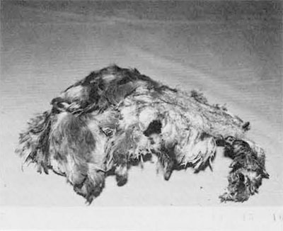
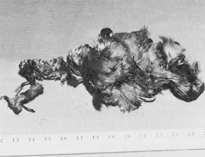
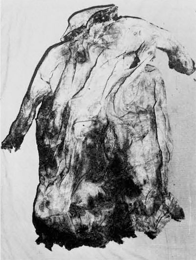
The mummy was unwrapped by removing five layers of sealskin and an innermost eiderdown parka. These summer parkas are customarily worn with the feathers outside, but this was apparently reversed for burial. The grave goods were limited to two bird skins, one a cap and the other, wedged between the arm and abdomen, an empty pouch, probably meant for carrying magical charms.
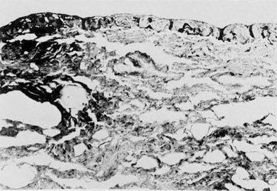
Apart from the fact that the tissues were dried and leather-like, necessitating the use of an electric saw, of the type used to remove plaster casts, rather than a scalpel, it was possible to conduct the autopsy in a relatively standard technique. The heart and lungs were well preserved but the abdominal viscera and brain had deteriorated markedly. The most striking gross pathological change was the appearance of the lower lobe of the right lung, which was reduced to a putty-like mass, in contrast to the remainder of the lung which showed preservation of the normal spongy architecture.
Microscopic examination confirmed the destruction of the right lung, revealing it to consist of clumps of bacteria of a specific type known as gram-negative bacilli. (Bacteria are generally divided into either bacilli, rodlike bodies, or cocci, which are round. They are also categorized, by their ability to take the gram stain, as gram-positive or -negative. The resultant four possible combinations all occur in nature and all can cause disease in man.) While many of the other organs showed post-mortem contamination by gram-positive and negative cocci, the heart, kidneys, and remainder of the lung contained discrete crystalline areas which contained the same gram-negative bacilli. This pattern is exactly what one would see in a modern patient with a bacterial pneumonia complicated by abscesses in other organs of the body.
The bacteria were no longer viable, as attempts to culture them were unsuccessful. The appearance of the bacteria in the tissue sections was distinctive enough that we were able to suggest that they were Klebsiella pneumoniae, an organism which causes a similar pneumonic infection in modern patients.
This person showed a number of other pathological changes. As in the Eskimo woman, anthracotic pigment had accumulated in his lungs, again because of indoor fires. Russian explorers had noted eye changes in the Aleuts due to their smoke-filled houses, and modern visitors have found it impossible to stay in Aleut houses for the same reason. He also showed some arthritis of the spine, an aging change to which Aleuts are particularly prone. The degree of arthritis was consistent with an age estimate of 30-50 years, an estimate supported by the finding of a moderate degree of arteriosclerosis.
There were a number of important negative findings, such as the absence of any evidence of physical trauma, poisoning, drowning, parasites, or tuberculosis, a disease presumed to be introduced by the Russians. Other special studies revealed him to be blood group 0, and to have marked dental attrition, due to severe masticatory stress, and periodontal disease, with heavy calculus formation.
The historical reconstruction of this person indicates that he was a man of modest status but with the unusual features for an Aleut of a mustache and beard. He died of lobar pneumonia, a disease which was commonly fatal until the use of the sulfa drugs in the 1930’s and penicillin in the 1940’s. He was simply wrapped in his summer parka and taken by boat to the burial cave on uninhabited Kagamil Island, where he was discovered by Hrdlicka some 200 years later.
This body serves as a visible reminder of one phase of the complex Aleut culture which was so abruptly disrupted by Western contact in the mid-18th century. This man showed no evidence of the epidemic diseases, tuberculosis, syphilis, smallpox, and measles, introduced by the Russians. These diseases were as much responsible for the deaths of many Aleuts as was the well-documented direct and violent conflict.
For most people the word mummy refers specifically to the embalmed and wrapped specimens of ancient Egypt. In predynastic times bodies buried in the hot sands of the western desert were noted to become quickly desiccated and were well preserved. By the beginning of dynastic times, some 5000 years ago, above-ground tombs were being built, allowing the bodies to decompose. The ancient Egyptians believed that the survival of the personality, or soul, depended on its ability to identify with the body; it therefore became necessary to develop an embalming technique.
The decomposition of tissues is due to the action of enzymes which break down proteins. Living cells have certain mechanisms to protect themselves against these enzymes, which are present in the cells themselves and in the bacteria which spread throughout the body after death. These enzymes can be inactivated either by chemical coagulation, the mechanism of modern embalming fluids, or by drying. The mummification technique of the Egyptians was an imitation of the drying of natural mummies, and was based on the use of natron, a sodium carbonate salt solution. The effect was to draw out water from the body, much the same as salting meat. The internal organs were removed and embalmed separately, except for the brain, which was discarded as of no value. The mummified organs were either replaced in the body or put in canopic jars to be stored in the tomb. In later periods the body was coated with bitumen, a pitch-like compound, in an attempt to hasten the mummification process by excluding air. An unfortunate result of this technique is that it makes the examination of such mummies extremely difficult.
A large number of Egyptian mummies have been examined since the mid-19th century. The major worker in the field was Sir Marc Armand Ruffer, an English physician who dissected hundreds of mummies in the early 20th century. Ruffer was the first to apply modern techniques to this material, and is considered the father of paleopathology. He discovered the antiquity of such processes as arteriosclerosis and schistosomiasis, a parasitic infestation which remains a serious problem in modern Egypt.
Since Ruffer’s untimely death in WWI, only sporadic reports on individual mummies have appeared in the literature. Recently a group has been brought together under the auspices of the University Museum, the Smithsonian Institution and the Paleopathology Association to begin a systematic study of the paleopathology of Egyptian mummies. Included are a number of pathologists and other physicians, archaeologists, physical anthropologists, biochemists and other scientists.
The first mummy examined by this group, in 1972 at the University Museum, was dubbed PUM-I (Pennsylvania University Museum). He seems to have been unable to afford proper embalming, and his mummy was somewhat of a disappointment, consisting of little more than a wrapped skeleton. Some of the organs were still in place, so he had not been eviscerated. Miscroscopic and chemical examination revealed severe bacterial decomposition. A touch of comic relief was provided by the finding of a 100-year-old railway ticket stuffed into a hole in the chest. (Someone must have felt that the mummy needed a ticket for its trip to the United States in the 19th century!)
Evidence of disease in this mummy was limited to an X-ray showing apparent tuberculosis, and a skin disorder which has only recently been described in modern patients. An interesting negative finding was the virtual absence of lead in the teeth, in contrast to the high levels in modern man.
Examination of PUM-II, at Wayne State University, in 1973, was much more satisfactory. This mummy, dated at 170 B.C., had been carefully preserved, so carefully, in fact, that it took six investigators a full day to remove the bitumen covering the body. It was found that the canopic parcels had been placed within the body. It was possible to identify the heart, major blood vessels, lungs, intestines, and spleen. As we have come to expect, the lungs contained a great deal of anthracotic pigment, but a surprise was the finding of considerable amounts of silica. This is presumed to be due to inhalation of silica-containing earth, and may indicate an occupational exposure.
PUM-II had arteriosclerosis, with large, fatty plaques involving the wall of the aorta, the major vessel leading from the heart. Other evidence of disease was the presence of an Ascaris Iumbricoides egg in the intestinal package. This is the earliest find of this common intestinal parasitic roundworm by over a thousand years.
An archaeological find of equal interest was that of a small piece of cleaned but unspun cotton in the mummy wrappings. This is the earliest cotton found in use in Egypt or, indeed, in the Western World, by some 500 years. Cotton is known to have been produced in India from at least 1500 B.C., and the question of whether this small piece was grown locally or imported is still under investigation.
As our paleopathology team includes a wide variety of scientific fields, many unusual facets of paleopathology can be explored. For example, we were able to examine PUM-II’s ears, and he was found to have perforated eardrums, evidence of the antiquity of middle ear disease. Blood typing and various chemical studies are also available to us. These techniques will be applied to the studies which are planned for the future, including an Egyptian mummy belonging to the Royal Ontario Museum in Toronto.
Of course, the best place to examine mummies is at the source, in Egypt. In the spring of 1974 I was fortunate enough to be invited to join the University Museum Expedition at Dira Abu el-Naga, on the west bank of the Nile, at the site of ancient Thebes. The modern city of Luxor, on the opposite east bank, is the site of the Egyptian capital during the first half of the New Kingdom. The Nile Valley at this point, 400 miles south of Cairo, is four to five miles wide, bounded on both east and west by limestone cliffs some 1000 feet high. The ancient Egyptians saw the sun setting on the west bank, beginning its nightly journey through the underworld to sunrise, so burials were on the west bank, for the soul of the deceased to follow the same path and rise again every day. The Theban necropolis contains the Valley of the Kings, burial place of the New Kingdom Pharaohs. Set into the eastern face of the nearby hills and cliffs are the tombs of hundreds of lesser officials, including three high priests of Amun, the chief god of Egypt, during the reign of Ramesses II (1304-1237 B.C.). The Museum Expedition is excavating these three tombs under the direction of Lanny Bell, as reported in Expedition in 1973. I examined the mummy and skeletal material removed from the tomb of the first of Ramesses’ high priests, Nebwenenef.
Two things were immediately apparent on surveying the 7500 specimens removed from the tomb. First, grave robbers had done a thorough job; 6000 of the specimens were fragments of bone too small to be identified and there were no whole mummies. Secondly, the tomb had been reused many times since Nebwenenef’s death about 1290 B.C. A head count (literally) revealed a minimum of 30 individuals, 25 adults and five children, to have been buried in the part of the tomb excavated to date. Several different styles of mummification were noted, indicating usage over many centuries.
One of the children was the closest specimen to an entire mummy, consisting of the trunk and arms. The desiccated organs were intact, and there was a prominent curvature of the spine, suggesting either tuberculosis or poliomyelitis, both of which have been documented in dynastic Egypt. Another possible cause, rickets, is very unlikely in Egypt’s sunny climate.
A disappointment was the absence of organ packages of the adult mummies. These probably had been destroyed by robbers looking for jewelry, just as they had destroyed all of the hands looking for rings. The bones showed very little pathology. An occasional example of periodontal disease or osteomyelitis was seen, and there were two examples of spina bifida, a congenital defect of the spine.
The specimens are en route to the Museum, and it is expected that microscopic examination will reveal more pathology. Among the specimens selected were the middle ears of all the heads, in an effort to determine the prevalence of middle ear disease as demonstrated in PUM-II. A number of teeth will be examined for lead levels, which, if they confirm the low levels found in PUM-I, will bolster our concept of lead accumulation as a result of modern pollution. Samples taken of blood vessels will be studied for arteriosclerosis, another presumably modern disease that actually appears to be of considerable antiquity.
There is a difficulty in studying these mummified tissues microscopically, that of separating the artefacts of drying and decomposition from ante-mortem pathology. The tissues must be rehydrated for study. Rehydration is based on a solution developed by Ruffer, consisting of water, alcohol, and sodium carbonate. This same solution is in use today, and accomplishes simultaneous rehydration and fixation of the mummified tissues. While the tissues can then be processed in the same fashion as fresh tissues, there are many changes in their microscopic appearance. The connective tissues, such as fat, muscle, and fibrous tissue are remarkably stable, but epithelial tissues fare poorly. This leads to major difficulties in diagnoses, as, for example, most tumors-are of epithelial origin. Foreign bodies such as bacteria, fungi, and worms are also very stable; bacteria have been reported from the tissues of a 5000-year-old Egyptian mummy.
As Director of the Autopsy Service at the Hospital of the University of Pennsylvania, I am able to approach this problem experimentally. Pathological specimens are removed from autopsy cases and desiccated in an oven, simulating the mummification undergone by bodies buried in the desert or artificially mummified by the application of natron. The specimens are then rehydrated and examined microscopically. The slides are compared to those made immediately after autopsy. Thus far the results have been most encouraging, in terms of diseases still being recognizable, but it must be remembered that the experimental setup represents almost ideal conditions. One can only expect true mummies to be less well preserved. However, such experiments do give information on which special techniques are useful in examining mummified tissue.
My plans for future investigations include the completion of the examination of the specimens from Dira Abu el-Naga, and the possibility of a return trip to Egypt, to examine a collection of intact mummies of known provenance. In conjunction with the members of the Paleopathology Association, the techniques of pathology, physical anthropology, Egyptology, microbiology, parasitology, physiology, radiology, electron microscopy and dentistry will be applied to these materials in an attempt to further delineate the role of disease in man’s physical, social and political history, and to aid in the understanding of the evolution of human disease.