A Complete View and a Complete Treatment: Conservation of the Roman Period Mummy Mask

Update – this post contains outdated language. We no longer use the term “mummy” and instead use “mummified human individuals” to refer to Ancient Egyptian people whose bodies were preserved for the afterlife. To read more about this decision, follow this link.   

After using humidification and four extra hands, the mask is now unfolded! This complete view of the object provides us a wonderful opportunity to look at the materials used in construction and allowed treatment to finally move forward.

Before jumping into treatment, I had the opportunity to perform Multispectral Imaging (MSI) on the mask, allowing us to analyze some of the pigments non-destructively and with great results.
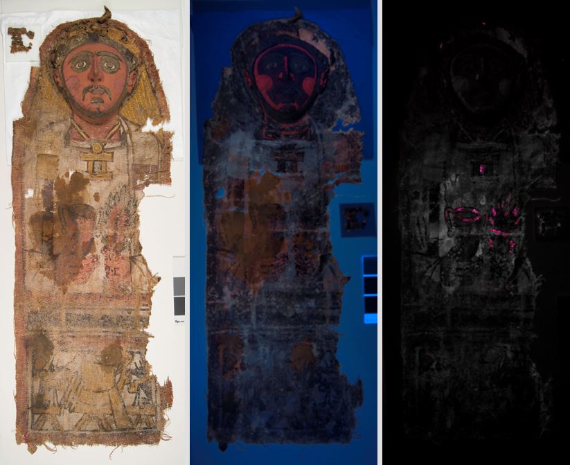
E2462. From left to right: Visible light, Ultraviolet illumination, Visible induced IR luminescence

E2462.
From left to right: Visible light, Ultraviolet illumination, Visible induced IR luminescence

Under ultraviolet illumination, a bright pink fluorescence was visible (middle), indicating the use of a madder lake pigment in the cheeks and to accentuate the face and hands. I also used visible induced IR luminescence to pinpoint the use of Egyptian Blue pigment in the crown, jewelry, and green leaves (right, Egyptian Blue highlighted in pink). This is a material commonly found in Roman period Egyptian artifacts.

In addition to finding out some of the materials used, I also completed full documentation of the object. Although some of the surface is still intact, the paint layer is in poor condition with areas of flaking and powdering. There is also a large loss to the textile along with some smaller tears and holes.

E2462 During treatment detail of flaking paint

E2462 During treatment detail of flaking paint

As my first order of business, the paint needed to be stabilized. This paint, like many other Egyptian painted surfaces, is sensitive to water and adhesives can cause staining and darkening. This meant a lot of testing was required to find the perfect adhesive for the job.

Using both testing panels and small, discrete areas of the surface, I tested adhesives until I found funori, a seaweed-based polysaccharide. This material preserved the matte and light tones of both the paint and ground layers.

Amaris Sturm, summer intern, consolidating surface of E2462

Amaris Sturm, summer intern, consolidating surface of E2462

As treatments usually go, you sometimes get unexpected bumps along the way. As I was consolidating I discovered that the flesh tones in the face and hands were significantly more sensitive to the water-based adhesive. I quickly had to rethink my approach, ultimately using a methyl cellulose in 50:50 ethanol: water for the hands, face, and larger flakes in the yellow framing the face.

Once consolidation was complete, I moved on to the next hurdle: the molded mud plaster mask. A large gap is present between the fragmented mud plaster crown and the textile below. To support the plaster and its mends, I made a removable fill of carved Volara foam and Japanese tissue, all toned with Golden acrylic paints to make the supports more discrete.

Removable fills to support the heavy mud plaster crown in E2462

Removable fills to support the heavy mud plaster crown in E2462

Fragmented, actively shifting, and detached mud plaster was mended with a 40% AYAT in acetone applied by brush and syringe. Unstable and weightbearing cracks and gaps were filled with a 25% AYAT in acetone that was bulked with microballoons and toned with dry pigments. Fill material was applied with syringed, shaped with a brush and wooden skewer, and  smoothed with a little bit of acetone. A thin toning layer of acrylic paint was applied to fills to make them a warmer tone, but still distinguishable from original material.

Filling compromised gaps on E2462

Filling compromised gaps on E2462

And with that, the treatment is complete! The mask is now stable and will be returned to storage safe and sound.

E2462 Before treatment (left) and After treatment (left)

E2462 Before treatment (left) and after treatment (right)

  • Amaris Sturm is a second-year graduate student in the Winterthur/ University of Delaware Program in Art Conservation. She recently completed her summer internship in the Penn Museum’s conservation labs.

New Mask in the Lab

Update – this post contains outdated language. We no longer use the term “mummy” and instead use “mummified human individuals” to refer to Ancient Egyptian people whose bodies were preserved for the afterlife. To read more about this decision, follow this link.   

Amaris Sturm is a second-year graduate student in the Winterthur/ University of Delaware Program in Art Conservation. She is currently completing a summer internship in the Penn Museum’s conservation labs.

I’m excited to introduce a new addition to the objects in the Artifact Lab! This Roman period Egyptian mummy mask and shroud, likely from 220 – 250 AD and excavated from Deir el-Bahri in the late 19th century, will be one of my primary treatment projects during my summer at the Penn Museum.

E2462- Overall before treatment

E2462- Overall before treatment

Meant to be placed over the upper body of a mummy, this mask is constructed of multiple pieces of coarsely woven linen sewn into a long shroud.  At the top of the shroud is a hollow, molded mud plaster mask in the form of a man’s face with a jeweled crown. The entire front surface has a white ground with colorful painted decoration. Additionally, gilding is present on fragments of the crown.

Sadly, the mask was folded at some point in its history, obscuring most of the linen shroud. Although there are no records of the complete decorated surface and little is known about the history of the mask in our collection, other similar examples from Deir el-Bahri give great insight into what may be hidden beneath the folds.

Comparable mask in the Louvre collection

Comparable mask from the Louvre collection

Comparable examples, including this mask from the Louvre, show the continuation of the man’s white tunic with a goblet in one hand and a plant stem in the other. A lower register is likely present containing Sokar, a falcon-headed god, on a boat and flanked by two jackals. One jackal is visible on an exposed corner of the Penn Museum’s mask.

E2462- Crown before treatment

E2462- Molded mud plaster crown before treatment

Apart from being folded, the mask has other condition issues that will be treated over the course of my summer internship. The textile support of the crown has sagged, causing the mud plaster to break and crumble. Additionally, the exposed painted surface is flaking and the linen fabric has started to tear and unravel.

I hope to start treatment in this coming week and unfold the shroud, allowing us to better understand the construction, decoration, and condition of this mummy mask. Check back to see what it revealed and for more on the mask’s treatment!

Sources:

Panel Portrait of a Man. Louvre Museum. Accessed June 25, 2016. http://www.louvre.fr/en/oeuvre-notices/panel-portrait-man

Riggs, C. 2000. Roman Period Mummy Masks from Deir el-Bahri. From The Journal of Egyptian Archaeology, 86. Egypt Exploration Society. 121-144.

A different sort of unwrapping…

by Alexis North, a project conservator spending the summer working with the Buddhist Murals Project, but who also has a strong interest in Egyptian materials. Read more about her work on Egyptian objects at the Michael C. Carlos Museum, Emory University, here.

Update – this post contains outdated language. We no longer use the term “mummy” and instead use “mummified human individuals” to refer to Ancient Egyptian people whose bodies were preserved for the afterlife. To read more about this decision, follow this link.

If any of you have visited the Artifact Lab in person, you may have heard us talk about how it was once popular to open or unwrap mummies, to see the body inside. Of course, this is no longer common practice, and we use non-invasive techniques such as x-radiography or CT scanning to see underneath a mummy’s wrapping without causing any damage or disturbance to the mummy’s current condition.

However, sometimes we are able to perform a slightly different kind of unwrapping, when items are found in storage in aging, opaque, or otherwise unsuitable housing conditions. Such was the case with this mystery item:

E12443, before opening and treatment

E12443, before opening and treatment

While it may look like Sunday’s dinner fresh from the butcher shop, it is actually supposed to be an ibis mummy. However, it has been wrapped in layers of tissue paper and plastic and you cannot see what the object actually looks like. While this type of storage is not damaging to the object, the fact that you cannot see the mummy inside makes this type of wrapping unsuitable. We always prefer to create storage supports or housings that allow researchers to easily see the objects without excessive handling. Therefore, this guy came up to the Artifact Lab for a little modern-day unwrapping.

E12443, after removing the plastic and tissue but before treatment

E12443, after removing the plastic and tissue but before treatment

And what a good-looking mummy it is! While we don’t have a lot of information about the age of this mummy, the intricate wrapping, which uses strips of both dyed and undyed linen, is typical of later periods in Egypt. It is also in very good condition, being just slightly dirty on the surface and having a few small areas of damage to the linen.

Detail images showing (1) a separated piece of linen wrapping on the top of the mummy, (2) a section of linen on the back torn and folded over, and (3) areas of loss which expose the ends of the woven linen underneath

Detail images showing (1) a separated piece of linen wrapping on the top of the mummy, (2) a section of linen on the back torn and folded over, and (3) areas of loss which expose the ends of the woven linen underneath

After gently cleaning the surface of the mummy using a vacuum and soft-bristled brush, I stabilized the areas of lifted or broken linen using Japanese tissue mends. Thin strips of tissue were toned brown using acrylic paint, then adhered underneath the lifting or broken areas using 2.5% methylcellulose adhesive in deionized water. I was able to reattach the broken piece of linen at the top of the mummy, and several sections of lifting wrappings which would be in danger of breaking, without stabilization.

I also humidified and reflattened the folded flap of linen on the back of the mummy. The opening caused by the folded flap was allowing fragments of the inner linen layers to break off and fall out. I used another Japanese tissue mend with methylcellulose to hold the reshaped flap in place.

Before (left) and after (right) flattening and readhering the flap of linen on the back of the ibis mummy

Before (left) and after (right) flattening and readhering the flap of linen on the back of the ibis mummy

Here are some images of the ibis mummy after I completed its treatment. I know it doesn’t look very different, and that happens a lot when treating archaeological objects. My goal wasn’t to improve or restore the mummy in any way, just make sure it could be safely handled and stored without any further damage.

    Images of (1) the top of the mummy, (2) the proper right side of the mummy, and (3) a detail of the reattached linen strip, after treatment

Images of (1) the top of the mummy, (2) the proper right side of the mummy, and (3) a detail of the reattached linen strip, after treatment

My last step was to make a new storage tray so the mummy can be easily seen and examined, without any wrappings besides the ones it came with!

The ibis mummy in its new storage mount

The ibis mummy in its new storage mount

Salvaging PUM I’s chest wrappings

This week, I started to work on the treatment of our mummy PUM I‘s linen wrappings. Poor PUM I – not only is his body quite deteriorated and in multiple pieces, but his linen wrappings are also fragmentary and very fragile. Some of linen in the worst condition are the pieces that once covered his chest, which were cut off during the 1972 autopsy.

This rectangular section of textiles was cut away as a single unit during the 1972 autopsy.

This rectangular section of textiles was cut away as a single unit during the 1972 autopsy.

In addition to the mechanical damage caused by the autopsy, the linen has suffered from insect damage and it is significantly stained and embrittled in areas, likely due in part to deterioration of the human remains they were once in contact with.

Removing the wrappings (left) and the chest wrappings after removal (right)

Removing the wrappings (left) and the chest wrappings after removal (right)

While this linen is in poor condition, it can be moved as a single unit, so we removed it for treatment. The goal of the current treatment is to keep the linen layers in this section together; to prevent them from slipping out of alignment and to prevent the linen from continuing to tear and deteriorate even more.

After vacuuming the linen thoroughly, I got to work relaxing distorted areas and realigning tears.

Local humidification of the linen in progress, using damp blotter and Gore-Tex

Local humidification of the linen in progress, using damp blotter and Gore-Tex

To realign tears, I bridged these areas from behind with small pieces of Japanese tissue paper, adhered in place with methylcellulose adhesive. The methylcellulose works well because it sets very quickly with only a small amount of pressure from my finger or a spatula.

One side of the wrappings before (left) and after (right) humidification and tear repair

One side of the wrappings before (left) and after (right) humidification and tear repair

The other side of the chest wrappings before (left) and after (right) tear repair

The other side of the chest wrappings before (left) and after (right) tear repair

This is only the beginning of the treatment on PUM I’s wrappings, but I think they are already looking better!


Giving our falcon a little love

One of my favorite artifacts in the lab is a falcon mummy, which I described in an earlier post. While he is a fascinating object, this poor little guy hasn’t been able to be exhibited, or even handled very much, because some of his linen wrappings are quite deteriorated, brittle, and breaking apart, causing serious structural issues.

Overall shot of our falcon mummy

Recently, I worked to stabilize the linen wrappings on his feet, which were partially detached, and in some areas, barely hanging on by a few threads.

Side view of the falcon’s “feet” showing the fragile, partially detached linen wrappings

Before carrying out any treatment, I did a little bit of research and carried out some testing to determine what materials I might want to use to repair the textile. I knew that a stitched repair would not be possible, as the linen fibers are far too weak and this would likely cause further damage, so I started investigating different adhesives and support materials to use instead. As part of this process, I consulted with Nancy Love, a local conservator in private practice who specializes in textiles. Nancy recently visited me in the Artifact Lab, and among the other materials I was trying, she suggested that I experiment with nylon bobbinett, a heatset nylon net.

I did some experimentation with it, and I really liked how it behaves, both as a support fabric and as an overlay to protect fragile areas-it drapes well and can be toned easily with dyes or paint. After feeling satisfied with the results of some of my tests, I set out to repair the damaged linen over the falcon’s feet.

I started by toning the bobbinett with Golden acrylic paint. Then I backed the fabric that was dangling off the back of the foot with the toned bobbinett lightly coated with 10% methylcellulose in water. I then used the bobbinett support fabric to raise the partially detached fabric up into place, secured temporarily with pins.

After positioning the linen, I covered the entire back of the foot area with another piece of toned nylon bobbinett.

The back of the foot area with an overlay of the toned nylon bobbinett, after treatment

Finally, I tacked down the strip of linen over the top of the feet, which was also partially detached but otherwise fairly stable, using small beads of methylcellulose. Reattaching the linen over this area also hid the edge of the nylon bobbinett overlay.

View of the front of the foot wrappings, after treatment

I’m pleased with the results, and I can now breathe a sigh of relief that we’re not going to lose any more of the linen from this area. My next task will be to address the falcon’s partially detached head/neck area. Hang in there, little guy!


Polarized Light Microscopy

Our Conservation Department recently purchased a Zeiss polarized light microscope-“the best microscope on campus” according to the specialist who set it up for us, and who is knowledgeable about the other scopes in use at Penn. Having the nicest equipment around isn’t familiar territory for conservation labs, so we’re enjoying having this status, but more importantly, having such a nice piece of equipment to use.

Our new microscope installed in the Artifact Lab

Polarized light microscopy (PLM) is used for examination of specimens in many types of laboratories, including biology and geology labs. In conservation, we use PLM for identification of minute fragments from objects-anything from pigment particles to wood fragments to textile fibers. We also use this technique to examine corrosion products, salts, and other materials found on artifacts-all of this work helps us better understand what the objects are made of, their condition, and ultimately provides important information for making conservation treatment decisions.

For example, our Conservation Fellow Tessa de Alarcon, who is conducting a year-long condition survey of Penn Museum artifacts from Kourion, Cyprus, has been using PLM to examine salts present in ceramic vessels from this collection. Tessa is desalinating the ceramics to remove the salts, which likely accumulated in the ceramics in the burial environment and will cause damage if not removed. To confirm which salts are present, she removed samples of the salts and examined them under the microscope. Here is an image of one of the salt samples, which shows that there are 2 different types of salts present-nitrates and sulfates.

Magnified image of 2 types of salts present on a ceramic vessel from Kourion (400X magnification).

You can read more about Tessa’s work with the Kourion collection (and view a cool video clip!) here on the Penn Museum blog.

In the Artifact Lab, one of the first ways that I’ve used our new microscope is to examine fibers from a thread that detached from the fabric wrappings of the falcon mummy I described in a previous blogpost. Fortunately for me (but unfortunately for the poor falcon mummy!) there are lots of detached threads that were available to sample for examination under the microscope. Here is a magnified image of one of these threads:

A small detached thread from the falcon mummy’s wrappings (40X magnification). I noted that the thread has an “S” twist and the fibers are shiny.

Using our binocular microscope, I put a drop of water on the thread and teased out several individual fibers from the thread on a glass slide, and then covered the fibers with a cover slip.

This image shows all of the tiny fibers from the larger thread-it is important to examine these fibers individually in order to identify what type of textile the falcon mummy is wrapped in (40X magnification).

Once the slide was prepared, I mounted it on the polarized light microscope and examined it at 50, 100 and 200X magnification.

Fiber from falcon mummy textile wrappings (200X magnification)

Under such powerful magnification, it is possible to see features such as a very small lumen (central cavity) and nodes along the length of the fiber. These features are characteristic of flax fibers, and comparing my sample with known references, it was immediately clear that this is what it is. Flax is used to make linen, and since the majority of ancient Egyptian textiles are linen, I already had a good idea that this is what was used to make the falcon mummy-but this proves it!

You can see from this work that PLM is a very useful technique, but it also is important to have an idea about what the possibilities are for what your sample-background research and close examination before microscopy is essential.